Paediatric Gastroenterologist | London | Hertfordshire | Online
Dr Daniel Crespi | Paediatric Gastroenterologist | Kids Gastro Care

Dr Daniel Crespi

Paediatric Gastroenterologist

Paediatric Gastroenterology Clinics
Elstree Hertfordshire 
Golders Green London
Video consultations available
Book Online


Specialist help for tummy troubles in children from newborn to 18

A Paediatric Gastroenterologist's guide to Diagnostic Tests used in paediatric gastroenterology

Helicobacter pylori test and other paediatric gastroenterology investigations used in children

When I discuss the concerns that you and your child have in one of my paediatric gastroenterology clinics, the clinical situation becomes clearer and often no further tests are needed.


In some circumstances though, and to help reach a diagnosis or exclude potential conditions, specialised tests can be used.  These add more information to the evolving picture. Any investigations are carefully chosen and tailored to your child's specific situation. 


There are various tests that are available to be used, including:


Blood tests - can be used to check for 

  • anaemia (when the body does not have enough healthy red blood cells and Haemoglobin - the protein that carries Oxygen to the body's organs)
  • inflammation - usually the body's way of protection from injury or infection when short-lived, but if chronic (long-lasting) can lead to damage to various organs (including the gut for example inflammatory bowel disease)
  • nutritional information including vitamin and mineral levels. 
  • kidney, liver and thyroid function
  • the immune system is intact and functioning well

as well as  to screen for and sometimes diagnose coeliac disease 


Stool or poo tests  - to check for infections, gut inflammation and biochemical analysis in times of malabsorbtion.


Imaging tests such as x-rays, ultrasounds or other scans including MRI can be used to look at the bowel in greater detail.  They can occasionally be helpful in estimating the gut transit time (gut motility studies) when difficult-to-treat constipation is a concern. Ultrasound and MRI scans can evaluate areas of the bowel or other digestive organs such as the gall bladder, liver, or pancreas for inflammation and the complications of inflammation.


Breath tests  -  can sometimes be used to check for carbohydrate intolerance such as Lactose or Fructose intolerance. 

They can also check for excessive bacteria in the small intestine (small intestinal bacterial overgrowth SIBO), which can cause a variety of gastrointestinal symptoms including bloating and change in bowel habit, some of which overlap with irritable bowel syndrome.

They rely on the principle that gases such as Hydrogen (and in some  Methane) are produced by bacteria in the bowel, that ferment poorly absorbed carbohydrates.  Some of the gas produced is absorbed and eventually exhaled and can be collected and analysed.


Paediatric Endoscopy and biopsy - can be used in some situations to look specifically at the lining layer (mucosa) of the digestive system in more detail.  


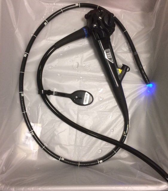
An endoscopy is a  procedure during which a trained endoscopist (e.g. a paediatric gastroenterologist, nurse endoscopist, adult gastroenterologist etc.) inserts an endoscope, a long thin floppy tube with a video camera and light at one end, into the digestive system via either the mouth or the bottom.  High-definition Images during the procedure are displayed on a monitor in front of the paediatric endoscopist so that the lining layer of the gut can be seen clearly. 


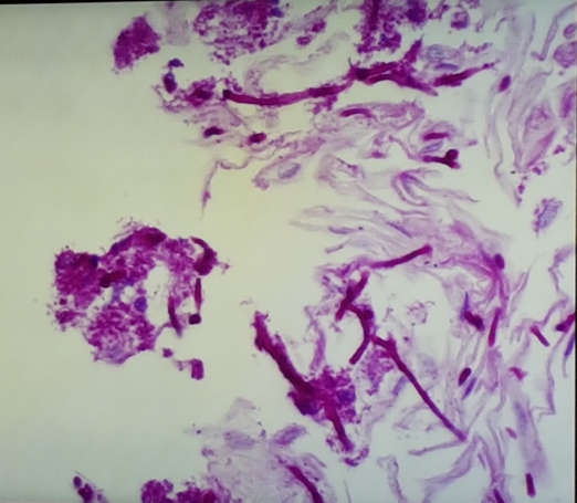
Whilst performing the procedure, Biopsy samples of tissue from the lining layer of the gastrointestinal (GI) tract are obtained for further detailed microscopic analysis by a pathologist. 


This can be useful when searching for the cause of symptoms such as pain and bleeding or concerns with growth, swallowing or vomiting. 


During the procedure, the naked eye of the endoscopist can see (macroscopic) changes such as ulcers or areas of redness that could possibly indicate inflammation.  The biopsies allow for further analysis under a microscope by a pathologist,  to check for the various types of inflammation involved, and for any  (microscopic) changes that are not always visible during the procedure.

 Paediatric Endoscopy V Adult endoscopy

In some circumstances, it may be necessary to look at the lining of the of the digestive system during a procedure known as an endoscopy. Specialised cameras known as endoscopes are used. These are long thin floppy tubes with a (video) camera and light source at one end that are inserted into the digestive system. Images are sent back to a high definition screen in front of the paediatric endoscopist. Whilst performing the procedure, biopsy samples from the lining of the gastrointestinal (GI) tract are obtained for further detailed microscopic analysis by a pathologist. 


Paediatric endoscopy differs from adult endoscopy in several ways hence the need for highly specialised training to be able to perform this test.


Some differences are that:

  • The equipment used is different for children. Although the inner workings of an endoscope are the same, paediatric gastroenterologists use different size gastroscopes and colonoscopes to take into account the different ages, size and weight of the children seen


  • some younger children are admitted overnight to have strong laxatives to prepare their bowels for colonoscopy whereas most adult colonoscopy is performed as a daycase visit to the endoscopy unit with bowel preparation given at home


  • The process of obtaining consent for procedures has some particular age related differences


Informed Consent for procedures


As for any significant procedure or test, before performing an endoscopy, one always needs to have the informed consent of the patient having the endoscopy. This is another aspect of paediatric gastroenterology and endoscopy that differs according to the age of the patient.

Close to the time of the procedure, a member of the team will meet with you and your child to discuss the benefits and potential risks of the procedure so that consent is a shared decision making process between the patient, family and doctor. 

A parent, or other adult with parental responsibility, must be present to sign the necessary consent form, in situations when a  younger child (younger than 16) needs to undergo endoscopy.


I have explained a great deal more about the different types of endoscopy in the FAQs section of the website, and for a child friendly video about what is involved, please have a look at the Useful Resources   page.  


My approach to your child is a personalised one.


I generally use the least invasive approach possible to work out a diagnosis and tailor it to your child's specific circumstances.


In some situations with a through history, clinical examination and completed bowel diary no tests are needed.

Sometimes, tests can be useful to confirm a clinical suspicion, or to rule out alternative possibilities that would otherwise need a different treatment strategy. For example when considering irritable bowel syndrome it is important to exclude coeliac disease.

It is important to remember that only If certain clinical indications are present, does it then becomes necessary to consider using more invasive investigative techniques such as endoscopy.

childrens endoscopy in London

Paediatric endoscopy 


The image shows a freshly disinfected and prepared endoscope still in its protective container. At its bendy tip one can see a light source that allows a well lit, clear view of the inner lining or mucosa of the digestive system to be transmitted to the high resolution monitor facing me during the procedure.  


The handle of the scope has two wheels that allow the endoscopist to control the position of the tip of the scope up, down, left and right. This allows for steering and fine movements of the tip of the scope.  


Below the wheels one can see the cap on the instrument channel - this is where a variety of instruments and equipment such as biopsy forceps, polypectomy snares, clips, and nets can be passed through the inner channels of the scope. The instrument then emerges at the tip of the scope within the bowel so that it can be used inside the bowel, as directed by the endoscopist.


Endoscopy is another area of difference between adult and paediatric gastroenterology.  The size of the patients I see can vary from an infant of just 10kg in weight, to a strapping 6ft plus rugby playing older teenager weighing 70Kg or more.  

This needs to be taken into account when choosing the most appropriate sized endoscopy equipment for your child.

oesophagitis in children

CASE STUDY 

Feeding difficulties, vomiting and a feeling of food getting stuck. 

Candida oesophagitis versus eosinophilic oesophagitis.


A recent case highlights the importance of using the right tests at the right time and how the accurate interpretation of results influences the correct choice of treatment.  


In this situation, the family and their GP were concerned about poor growth and weight gain as well as vomiting from an early age.  They had had various imaging tests performed to look at the structures of the upper gastrointestinal tract that had been normal.  Because they also had experienced problems with swallowing their food it was suspected that they may have inflammation affecting their foodpipe. For this reason a decision was made to proceed with an upper GI endoscopy to look for a particular type of allergic inflammation known as eosinophilic oesophagitis. 


The image here is that of a biopsy from the lining of the oesophagus or foodpipe.  The samples are carefully handled and stored in special containers whilst being transported to the laboratory where they are then processed with specific stains. These highlight certain features of the cellular structure of the region being examined.  

Here one can see several long filaments or hyphae (deep purple stain) of a fungus known as Candida Albicans, more commonly known as the fungus that causes thrush.  It can contribute to a variety of symptoms including vomiting and  difficulties with swallowing such as pain. 

This infection is occasionally found in the mouth and oesophagus especially in those using steroid medications given via inhaler, particularly if the incorrect technique is used. 

During the endoscopy one can see white plaques of material on the lining of the oesophagus.  


The appearance of Candida during an upper GI endoscopy can be similar to that seen in  another condition known as eosinophilic oesophagitis (EoE). This is a very different type of problem where there is allergic inflammation affecting the oesophagus.   


The biopsy findings, as well as special micobiology tests aiming to grow the fungus, helped to prove the diagnosis. 


The treatments of the two conditions are very different. One requires treatment with anti-fungal medication, and the other often needs dietary changes to be made.


In eosinophilic oesophagitis, a type of white cell known as the eosinophil, is found in the lining of the food pipe in biopsies collected during an upper GI endosocpy.  These cells are the same type of cell found in certain allergy related  conditions such as asthma, eczema or hayfever.  


Eosinophils are part of the body's immunological defence system as they protect against parasite infections. It is thought that they are effective against parasites, as they contain small granules or tiny packets, within which are chemicals that when  released can wreak havoc and cause damage to the invading parasite.  


However when these cells are found in areas of the body and are activated in the absence of any parasites, the damage is inflicted on the surrounding organ such as the foodpipe (eosinophilic oesophagitis), lung (asthma) or skin (eczema). The ensuing symptoms are related to the organ involved.


Share by: